All adults should have a sight test and eye health check every two years. If you have an eye condition, an optometrist at your opticians may recommend that you have more frequent checks.
Some optometrists carry out sight tests and eye health checks in people's homes. Ask your opticians if they offer this service for patients who are unable to leave their homes alone.
Many people are entitled to get free NHS sight test or optical vouchers to reduce the cost of their glasses or contact lenses. Find out if you're entitled to a free NHS sight test or an optical voucher on this page Free NHS eye tests.
Below are some of the other eye tests that you may need if you have a problem with your eyes or eyesight.
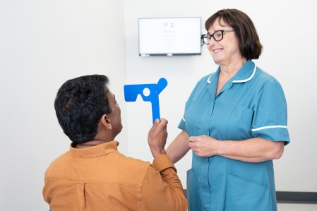
These tests measure uncorrected vision which is how well you can see without your glasses or contact lenses. They can also measure the best corrected vision which is the best possible vision you can achieve with glasses or contact lenses. If you are unable to read or recognise letters, alternative tests can be used with images. These tests are often used to test visual acuity in young children.
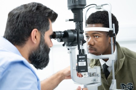
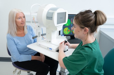
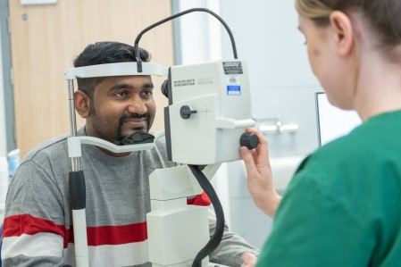
The eye care professional uses a special microscope to shine a beam of light, shaped like a small slit, into your eye.
Eye drops may be used to dilate your eyes first.
This test measures your peripheral (non-central) vision. You’ll stare at a target in the centre of your line of vision. As you look at that target, you’ll click a button when you see an object moving into your field of vision or, depending on the test, when a dot of light appears. This test lets the eye care professional know if conditions like stroke or glaucoma have damaged areas in your vision.
This video produced by the Royal Free London NHS Foundation Trust explains what is involved in having a visual fields test:
Refraction tests are used to get the prescription for your glasses. You look through a device called a phoropter at a chart about 6 metres (20 feet) away, or in a mirror that makes things look that distance away.
The eye care professional will then move lenses of different strengths in front of your eyes so you can tell them if things look clear or blurry. Your answers will give them the prescription for your glasses or contact lenses. The test will also help the eye care professional spot other eye conditions. Nothing will touch your eye so you won’t feel any pain or discomfort.
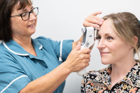
There are two different types of tonometry tests.
Applanation tonometry measures the amount of pressure it takes to flatten a portion of your cornea, the clear front part of the eye. You’ll be given drops to numb your eye, then a tonometer will be pressed lightly on it to measure the pressure.
Non-contact tonometry is often called the ‘puff of air test’. A different tonometer blows a tiny puff of air onto your eye. This measures eye pressure indirectly by the eye's resistance to the puff of air.
There is more information about the tests for diagnosing glaucoma on the NHS website. The World Glaucoma Association also has information about tonometry on the WGA website.
These tests check the function and performance of your visual system which includes your eyes, connecting pathways and the parts of your brain that deal with sight. Sensors are attached to the skin at the back of your head or around your eyes to pick up tiny electrical signals. These signals happen when your visual system is stimulated, either by patterns on a TV screen or by a flashing light. The tests are painless and take around 30 minutes.
This animation explains what to expect when you go for an electrodiagnostic test:
A gonioscopy test looks at the ‘angle’ of your eye. This is the fluid-filled space between the iris, the coloured part of your eye, and the cornea, the clear front part of your eye. The angle is where the fluid should drain out of your eye. A gonioscopy can check if this area is open or blocked. If it is blocked, it can affect how fluid drains out of your eye.
For the test, your eyes will be numbed with eye drops. The eye care professional will then place a special contact lens with mirrors directly on your eye and shine a beam of light into the lens. You may feel the lens touch your eyelashes, but gonioscopy is not painful at all. The test usually takes just a few minutes.
The World Glaucoma Association has more information about gonioscopy on its website.
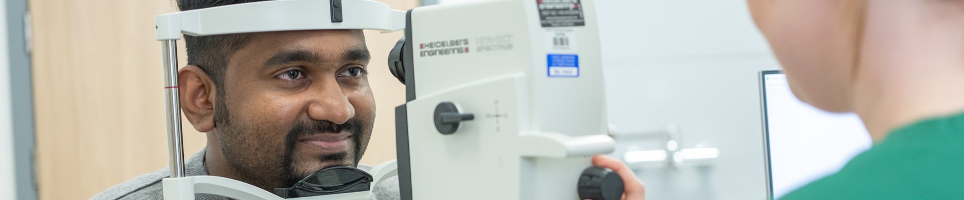
Corneal tomography produces a 3-D image of the cornea. It does this by mapping out the entire cornea using a camera that captures cross-sections of the cornea as it rotates. Like corneal topography, it is painless as nothing touches the eye.
There is more information about corneal topography on the All About Vision website.
Ocular biometry measures the size of the eyeball to work out the prescription needed for the new lens that replaces the eye’s natural lens during cataract, laser eye surgery and lens surgery. You will sit at a machine and be asked to look at a light. It is another painless test as nothing touches the eye.
There is more information about optical coherence tomography on the Cardiff University Eye Clinic website. You can find detailed information about the OCT scan on the College of Optometrists website.
This type of photography uses special cameras that can see your retina, the light-sensitive area at the back of your eye. Filters can be used to show your eye in different ways to help the eye care professional diagnose or check your condition.
Some retinal cameras can photograph a larger area of your retina to give the eye care professional a better view of the back of your eye. These ultra-wide cameras are ideal for conditions such as diabetic retinopathy. This condition can appear in parts of your peripheral (non-central) retina, as well as the central part you use for your central vision.
For this simple test, you place your chin on a chin rest and you’ll see flashing lights as the photographs are taken. Nothing will touch your eye and the test is painless.
There is more information about retinal photography on the All About Vision website.
Angiography is a type of test used to check blood vessels. Fluorescein angiography (FA) and indocyanine green (ICG) angiography help diagnose conditions like diabetic retinopathy, retinal detachment, and macular degeneration. Fluorescein and indocyanine green are the names of special dyes used to highlight blood vessels in the eye.
If you have an angiogram, an eye care professional will inject one of these dyes into a vein in your arm. The dye travels quickly to the blood vessels inside your eye. The eye care professional will then use a retinal camera to take photographs with special filters to highlight the dye. This helps them see any circulation problems, swelling, leaking or if there are any abnormal blood vessels.
These scans use sound waves to produce a picture of the inside of the eye. It helps your eye care professional diagnose and treat tumours, cataracts or bleeding in your eye. You might also get an ultrasound before cataract surgery. The scan only takes a few minutes and it is painless. Much like an ultrasound scan performed elsewhere on the body, a probe is smeared with a liquid gel and moved over the closed eyelid. The information is captured on a computer which makes images of what the inside of your eye looks like.
There is more information about ultrasound scans on the Moorfields Eye Hospital website.